The airport x ray machine images comes equipped with advanced digital detectors that transform X-ray energy into high definition images of incredible detail. The system's design makes it easy to use and facilitates quick image capturing. The airport x ray machine images system can be connected effortlessly to hospital information systems that enable the secure transfer of data. The system's robust design provides support for long-term use within healthcare settings.

Apart from traditional diagnostics, the airport x ray machine images is applied in interventional procedures to assist physicians in minimally invasive treatments. It provides real-time imaging during catheter placement, spinal manipulations, and orthopedic implant confirmation. The airport x ray machine images enhances the accuracy of the procedure and patient safety in complex medical procedures.
With advancements in technology, the airport x ray machine images will get progressively smaller, intelligent, and networked. It will also support augmented reality for training and procedural guidance. The airport x ray machine images will have self-calibration and automated maintenance functionality, which will increase reliability and operational performance.
The life of the airport x ray machine images relies on proper maintenance and surveillance. The X-ray tube, generator, and control panel are some of the parts that need to be examined and serviced based on manufacturer recommendations. The airport x ray machine images should be protected from moisture, vibration, and heavy dust to prevent performance loss.
In today's healthcare system, the airport x ray machine images continues to be an integral part of diagnostic imaging. The airport x ray machine images provides precise visual data that helps in disease detection and assessment of an injury. The airport x ray machine images has digital sensors and the capability to improve images. The airport x ray machine images helps in quick and effective medical imaging.
Q: What makes an x-ray machine different from a CT scanner? A: An x-ray machine captures a single 2D image, while a CT scanner takes multiple x-rays from different angles to create 3D cross-sectional views. Q: How is image quality measured in an x-ray machine? A: Image quality depends on factors like contrast, resolution, and exposure settings, which are adjusted based on the target area being examined. Q: What power supply does an x-ray machine require? A: Most x-ray machines operate on high-voltage power systems, typically between 40 to 150 kilovolts, depending on their intended use. Q: Can x-ray machines be used for dental imaging? A: Yes, specialized dental x-ray machines provide detailed images of teeth, jaws, and surrounding structures to support oral health assessments. Q: How does digital imaging improve x-ray efficiency? A: Digital systems allow instant image preview, faster diagnosis, and reduced need for retakes, improving workflow efficiency in clinical environments.
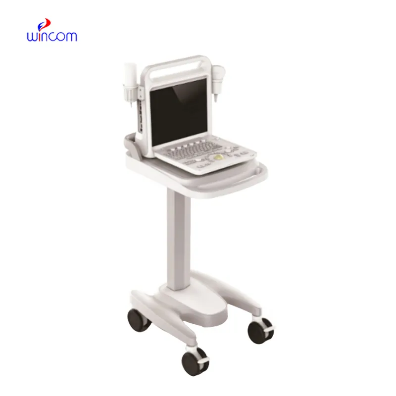
This ultrasound scanner has truly improved our workflow. The image resolution and portability make it a great addition to our clinic.
The microscope delivers incredibly sharp images and precise focusing. It’s perfect for both professional lab work and educational use.
To protect the privacy of our buyers, only public service email domains like Gmail, Yahoo, and MSN will be displayed. Additionally, only a limited portion of the inquiry content will be shown.
We are planning to upgrade our imaging department and would like more information on your mri machin...
I’d like to inquire about your x-ray machine models. Could you provide the technical datasheet, wa...
E-mail: [email protected]
Tel: +86-731-84176622
+86-731-84136655
Address: Rm.1507,Xinsancheng Plaza. No.58, Renmin Road(E),Changsha,Hunan,China

 af
af
 es
es
 ar
ar
 tr
tr
 sw
sw
 pt
pt
 th
th
 ur
ur
 bn
bn
 ne
ne
 vi
vi
 km
km
 lo
lo
 de
de
 ru
ru
 fi
fi
 nl
nl
 fa
fa
 fr
fr
 ko
ko