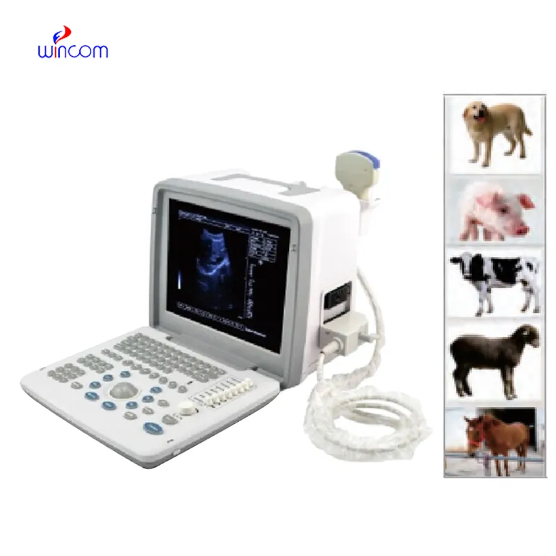
Through a combination of novel imaging software and the cow ultrasound scanner, the professionals can be very precise with the details of the anatomy they have captured and even more so with the accuracy of the final result. An ergonomic design of the device contributes to the reduction of operator fatigue during long use. The device, the cow ultrasound scanner, also comes with the ability to connect to more than one probe thus giving it the flexibility required when catering to various diagnostic needs. Besides that, the data export and connection functions of the device help to decrease the time taken in report-generating which is done through image sharing.

In medical imaging, the cow ultrasound scanner is a trustworthy device for different clinical departments. It provides vascular studies support by observing the state of the arteries and veins; in urology, it has a role in the assessment of the bladder and prostate health. The cow ultrasound scanner also enables emergency medical staff to make quick trauma evaluations, directing their actions precisely and efficiently.

The coming years will see the evolution of the cow ultrasound scanner into an independent and adaptable imaging solution. The increased level of automation will eliminate the need for human input. The cow ultrasound scanner may include predictive model components that will help healthcare providers to identify probable risks to an individual's health.
In order to extend the service life of the cow ultrasound scanner, it is recommended that users refrain from applying much force during the process of connecting/disconnecting probes. Power cables should always remain dry. The cow ultrasound scanner needs diagnostic tests to ensure that it produces quality images.
The cow ultrasound scanner represents an advanced form of medical imaging technology that transforms sound waves into high-definition visual data. It is widely used for evaluating organ health, tracking fetal development, and detecting vascular conditions. The cow ultrasound scanner ensures real-time monitoring and fast diagnostic results, supporting effective clinical workflows.
Q: What imaging modes are available on the ultrasound scannert? A: It supports multiple modes such as B-mode, M-mode, and color Doppler for diverse diagnostic applications. Q: How does the ultrasound scannert improve diagnostic accuracy? A: By providing high-resolution images and real-time feedback, it enables more precise medical evaluations. Q: Can the ultrasound scannert be used in field or remote settings? A: Yes, its portable versions are designed for mobility and can be used in clinics, hospitals, or mobile healthcare units. Q: What kind of display does the ultrasound scannert use? A: It typically features a high-definition digital display that enhances image visualization and readability. Q: How is data from the ultrasound scannert managed? A: The device allows secure storage, easy access, and export of imaging data through USB or network connections.
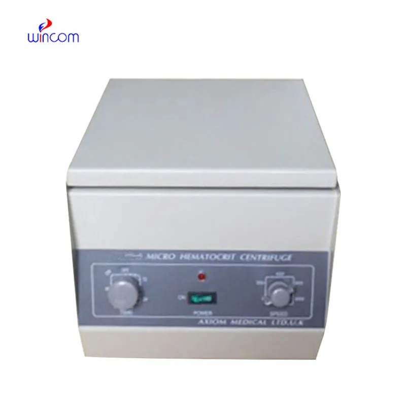
We’ve used this centrifuge for several months now, and it has performed consistently well. The speed control and balance are excellent.
The centrifuge operates quietly and efficiently. It’s compact but surprisingly powerful, making it perfect for daily lab use.
To protect the privacy of our buyers, only public service email domains like Gmail, Yahoo, and MSN will be displayed. Additionally, only a limited portion of the inquiry content will be shown.
We are planning to upgrade our imaging department and would like more information on your mri machin...
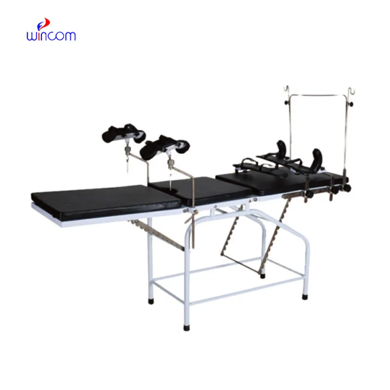
Could you share the specifications and price for your hospital bed models? We’re looking for adjus...
E-mail: [email protected]
Tel: +86-731-84176622
+86-731-84136655
Address: Rm.1507,Xinsancheng Plaza. No.58, Renmin Road(E),Changsha,Hunan,China

 af
af
 es
es
 ar
ar
 tr
tr
 sw
sw
 pt
pt
 th
th
 ur
ur
 bn
bn
 ne
ne
 vi
vi
 km
km
 lo
lo
 de
de
 ru
ru
 fi
fi
 nl
nl
 fa
fa
 fr
fr
 ko
ko