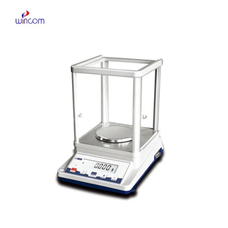
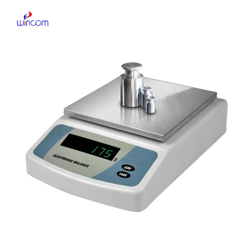
electronic balances is very important for laboratories that need to be very precise in their measurements of tiny amounts, for example, in clinical, pharmaceutical and research environments. It also helps very well in the accurate preparation of reagents, solutions and samples for analysis. The laboratory staff depends on electronic balances for keeping the same quality, method validation and getting accurate results. Its sensitivity, dependability, and reproducibility make it a necessary tool not only for hospital laboratories but also for research institutes and pharmaceutical quality control processes.
Hospitals' analytical laboratories use electronic balances during the production of internal standards for instrument testing methods. Very accurate mass input is a must to maintain uniformity over different analytical runs. This use case allows for comparison of still data, traceability, and monitoring of analytical performance over a long period. By allowing exact preparation of reference materials, electronic balances boosts measurement confidence in all phases of hospital laboratory work.
The ongoing evolution of regulations will see the electronic balances adding features for compliance support that will be more advanced. Hospital lab audits and accreditation standards will be met with the help of the documentation functions and secure data storage. This future trend will greatly improve the quality management processes in all hospitals and research labs.
In order to keep electronic balances in a good condition consistent calibration practices are needed that follow hospital laboratory protocols. Scheduled calibration checks are performed to maintain the reliability of measurements during daily activities involving analysis. Conditions in the environment such as temperature and the amount of air that moves around should be kept under control so as to prevent drift. The people operating the machines should make sure that there are no sudden changes in load and that the weighing pan is not subjected to excessive force. Through adhering to controlled handling practices, electronic balances is always trusted for pharmaceutical preparation and medical research activities.
Balance is crucial in the various ranges of hospital and clinical laboratories for the preparation of patient samples to be analyzed. Because weighing correctly provides proper reagent ratios, it ensures consistent dilutions and valid diagnostic test results. Laboratory staff can achieve a huge array of quality standards in sample preparation with electronic balances, being assured of reliable clinical diagnostics, treatment monitoring, and patient safety by means of precise measurement of laboratory materials.
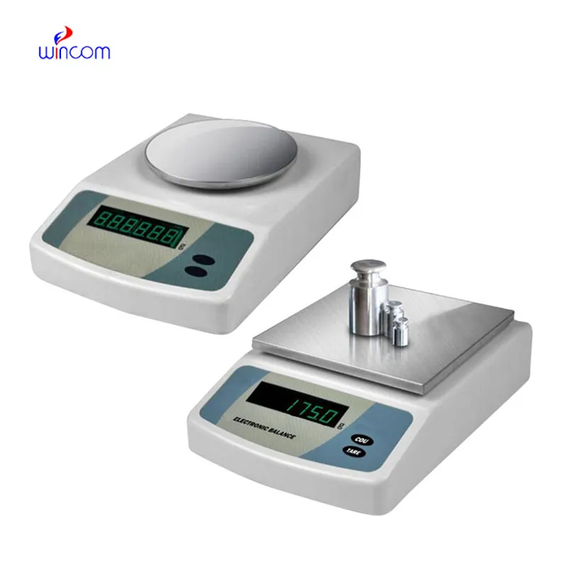
Q: What is the main purpose of an Analytical Balance? A: Its purpose is mainly to measure very tiny sample masses with the utmost precision in laboratories and hospitals. Q: What is the typical weighing range of an Analytical Balance? A: The weighing range for the majority of analytical balances is from 0 up to some grams with a resolution of micrograms or milligrams. Q: What environmental controls are necessary for an Analytical Balance's operation? A: Airflow, vibration, and temperature changes should not only be avoided but also prevented in the room where the scale is situated. Q: Is an Analytical Balance permitted in a hospital laboratory? A: Yes, it has indeed found widespread usage for the preparation of reagents, calibra¬tion, and drug development applications. Q: What should be the frequency of calibration for an Analytical Balance? A: The calibration interval is subject to the degree of use and the particular laboratory requirements.
This x-ray machine is reliable and easy to operate. Our technicians appreciate how quickly it processes scans, saving valuable time during busy patient hours.
The hospital bed is well-designed and very practical. Patients find it comfortable, and nurses appreciate how simple it is to operate.
To protect the privacy of our buyers, only public service email domains like Gmail, Yahoo, and MSN will be displayed. Additionally, only a limited portion of the inquiry content will be shown.
We’re interested in your delivery bed for our maternity department. Please send detailed specifica...
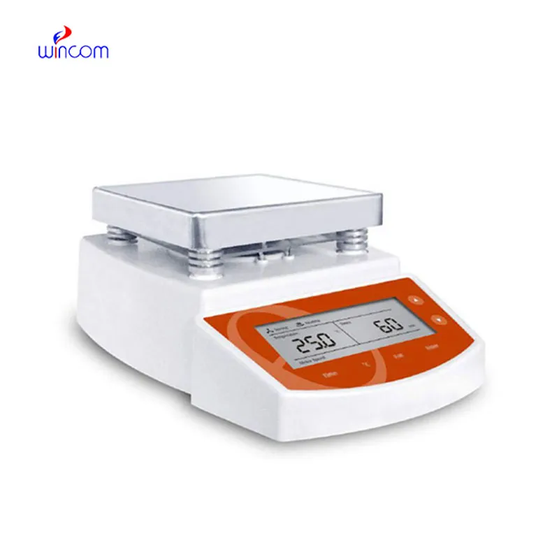
Hello, I’m interested in your water bath for laboratory applications. Can you confirm the temperat...
E-mail: [email protected]
Tel: +86-731-84176622
+86-731-84136655
Address: Rm.1507,Xinsancheng Plaza. No.58, Renmin Road(E),Changsha,Hunan,China

 af
af
 es
es
 ar
ar
 tr
tr
 sw
sw
 pt
pt
 th
th
 ur
ur
 bn
bn
 ne
ne
 vi
vi
 km
km
 lo
lo
 de
de
 ru
ru
 fi
fi
 nl
nl
 fa
fa
 fr
fr
 ko
ko