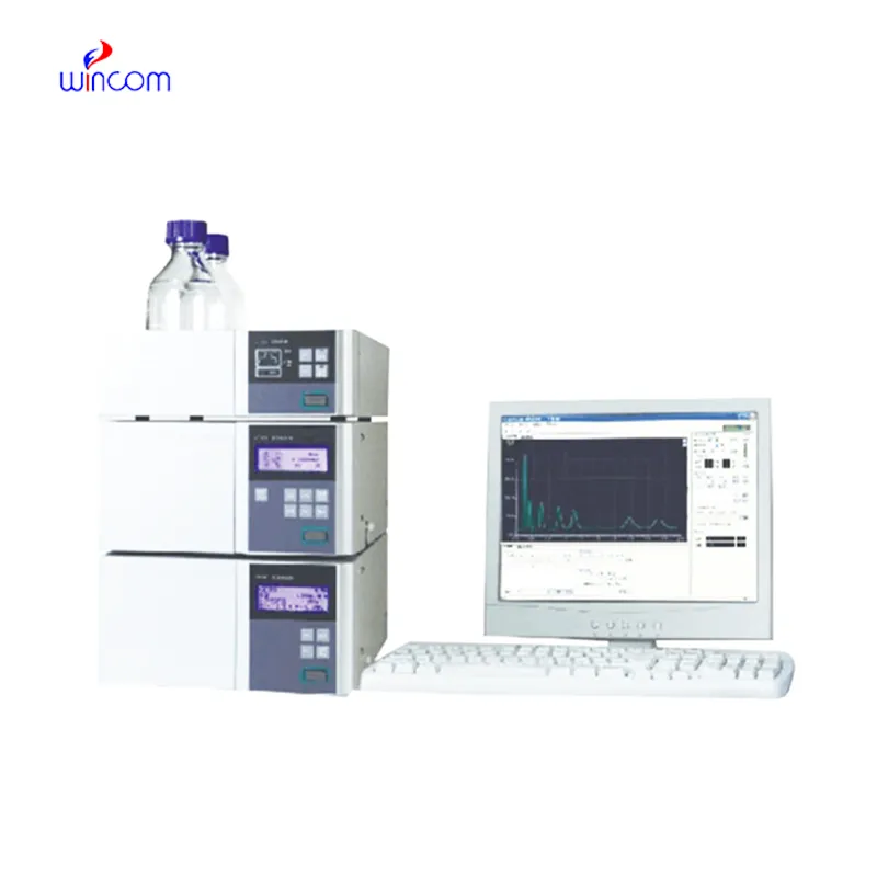
lc liquid chromatography is a critical technique to obtain analytical information in studies of medicines, clinical samples, and biochemistry. It isolates compounds according to their chemical characteristics, generating reproducible analytical results. Laboratory scientists use lc liquid chromatography to perform drug stability tests, monitor patient biomarkers, and find impurities. Its very high accuracy and flexibility allow thorough sample analysis in research, hospital, and clinical laboratory environments, thus becoming a fundamental device for assuring precision in both experimental and diagnostic results.
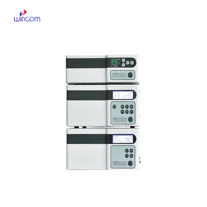
lc liquid chromatography are utilized by clinical laboratories for hormone and endocrine-related biomarker detection. It delivers trustworthy information for the diagnosis of endocrine diseases by correctly separating substances like cortisol, thyroid hormones, or insulin. Techs in a laboratory rely on lc liquid chromatography to give accurate and repeatable results, thereby helping doctors in individual treatment plan.
lc liquid chromatography is expected to have an increasing role in personalized medicine, analyzing complicated biomarkers swiftly. In the future, their application in hospitals will be centered on integrating pharmacokinetics, metabolomics, and monitoring, helping medical practitioners have access to swift and comprehensive data. The workflow in laboratories is expected to be organized.
lc liquid chromatography will require regular maintenance to be kept up in order to continue providing precise measurements in medical laboratories. After every use, the technicians should flush the columns, check the seals, and inspect the tubing for wear and tear and ensure that the detector is working. Regular calibration and good solvent management decrease the chances of system damage and increase the consistency of the results. Good care and maintenance not only increase the efficiency of the laboratory but also help in providing reliable diagnostics and maintaining the instruments for hospital applications.
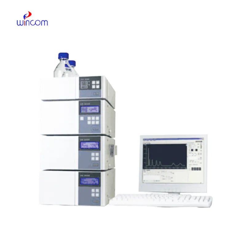
Clinical laboratories make use of lc liquid chromatography to analyze patient samples with remarkable accuracy. It identifies biomarkers, metabolites, and the levels of therapeutic drugs, thus giving reliable information about the disease status and monitoring treatment. Sensitivity of the technique permits determination of compounds in very minute amounts, which is critical in clinical testing. By resolving complex composition, lc liquid chromatography guarantees accurate and reproducible results for laboratory diagnostics. Lab staff utilizes it for daily testing, quality control, and research activities, thus making lc liquid chromatography a vital part of contemporary clinical laboratory work that caters to patient care, treatment choices, and lab data integrity.
Q: What is HPLC used for in laboratories? A: HPLC turns out to be one of the most significant and essential analytical methods in laboratories equipped with the chemical compound analysis, separation, identification, and quantification of their presence in complex samples which are the research, clinical, and pharmaceutical applications. Q: How does HPLC separate compounds? A: The HPLC separation technique is based on the different affinities of the compounds to the stationary phase and mobile phase within the chromatography column. Q: Can HPLC analyze biological samples? A: Yes, it is certainly possible to carry out analyses on various biological fluids such as blood, serum, urine, etc. for the detection of metabolites, drugs, and biomarkers. Q: How often should HPLC columns be replaced? A: The replacement of the columns must be done according to the manufacturer instructions or when the performance begins to decline, which is quite usual after heavy use or contamination. Q: What detectors can be used with HPLC? A: The analysis type determines the use of, among others, UV, fluorescence, refractive index, and mass spectrometry detectors as the common detectors.
We’ve used this centrifuge for several months now, and it has performed consistently well. The speed control and balance are excellent.
The delivery bed is well-designed and reliable. Our staff finds it simple to operate, and patients feel comfortable using it.
To protect the privacy of our buyers, only public service email domains like Gmail, Yahoo, and MSN will be displayed. Additionally, only a limited portion of the inquiry content will be shown.
Hello, I’m interested in your water bath for laboratory applications. Can you confirm the temperat...
I’m looking to purchase several microscopes for a research lab. Please let me know the price list ...
E-mail: [email protected]
Tel: +86-731-84176622
+86-731-84136655
Address: Rm.1507,Xinsancheng Plaza. No.58, Renmin Road(E),Changsha,Hunan,China

 af
af
 es
es
 ar
ar
 tr
tr
 sw
sw
 pt
pt
 th
th
 ur
ur
 bn
bn
 ne
ne
 vi
vi
 km
km
 lo
lo
 de
de
 ru
ru
 fi
fi
 nl
nl
 fa
fa
 fr
fr
 ko
ko