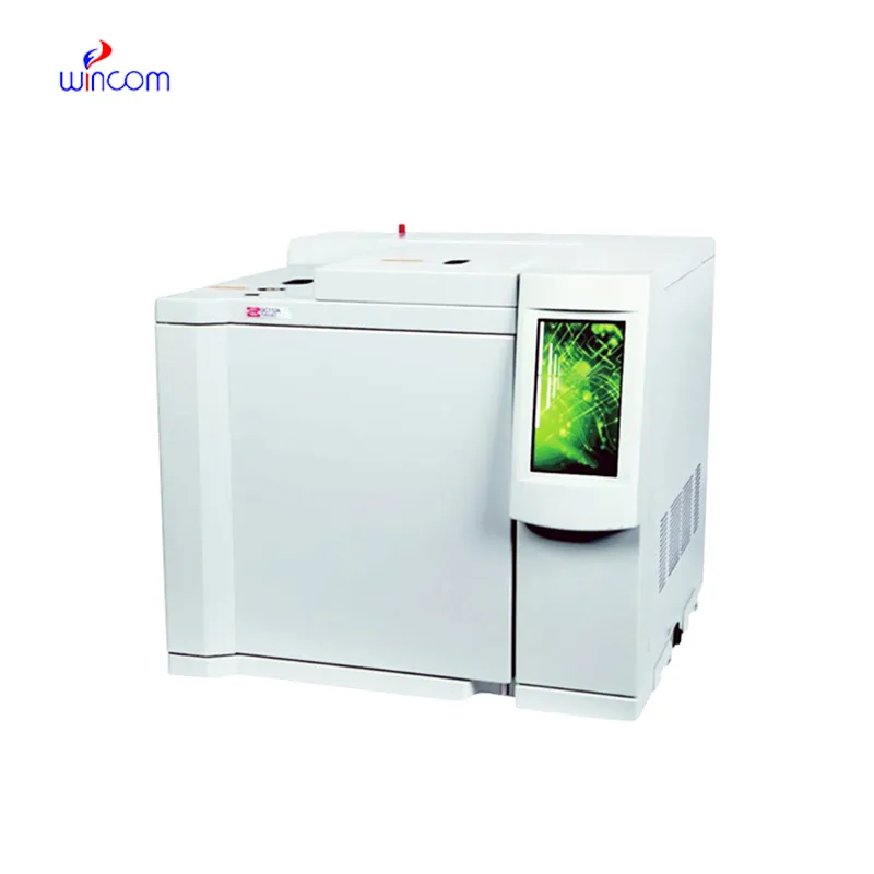
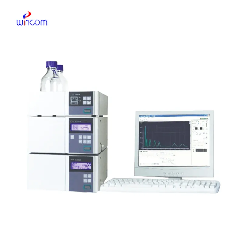
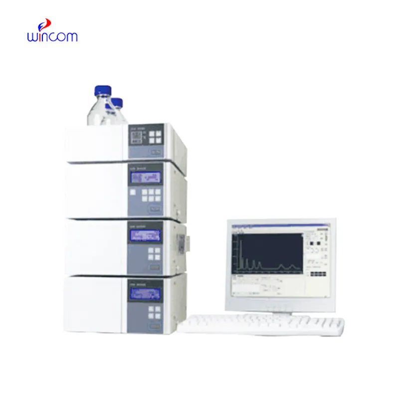
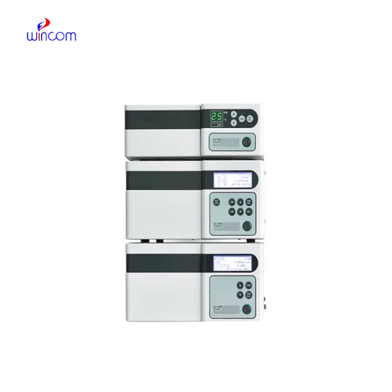
liquid chromatography mass spectrometry analysis is a primary tool in hospital and laboratory analytics. Its skills of isolating, measuring, and characterizing both chemical and biological substances enhance research as well as clinical testing. Quality control, drug testing, and testing of samples are done by laboratory technicians using liquid chromatography mass spectrometry analysis. The device's flexibility and reliability guarantee uniform performance, yielding critical analytical data that are vital for patient care, experimental validation, and smooth and fast laboratory operations in both healthcare and scientific domains.
liquid chromatography mass spectrometry analysis finds extensive application in hospital laboratories for monitoring drugs therapeutically. It provides precise determination of drug levels in patients' samples, thus making safe and effective dosing possible. Metabolites are tracked, treatment progress is assessed, and unexpected drug interactions are detected by the laboratory personnel. Its high accuracy and reproducibility facilitate both medical decision-making and research, hence, liquid chromatography mass spectrometry analysis becomes an indispensable instrument in taking care of patients and analyzing the medical field.
The future of liquid chromatography mass spectrometry analysis stresses the integration of hospital information systems and electronic medical records. The analysis of patient samples will be automatically included in the clinical workflows. Increased automation, AI-based interpretation, and better sensitivity will put liquid chromatography mass spectrometry analysis at the center of the laboratory operations and patient care that is focused on the patient's needs.
Systematic cleaning, pressure monitoring, and timely worn parts replacement are among the measures to be taken in the hospital laboratories to keep liquid chromatography mass spectrometry analysis under control. Laboratory staff must ensure the observance of the suggested operating conditions, avoid the formation of air bubbles in the system, and check for proper solvent compatibility. Regular maintenance maintains the performance of the column, avoids contamination, and allows the analysis to be precise and reproducible, thereby benefiting not only routine patient testing but also experimental research.
Clinical laboratories make use of liquid chromatography mass spectrometry analysis to analyze patient samples with remarkable accuracy. It identifies biomarkers, metabolites, and the levels of therapeutic drugs, thus giving reliable information about the disease status and monitoring treatment. Sensitivity of the technique permits determination of compounds in very minute amounts, which is critical in clinical testing. By resolving complex composition, liquid chromatography mass spectrometry analysis guarantees accurate and reproducible results for laboratory diagnostics. Lab staff utilizes it for daily testing, quality control, and research activities, thus making liquid chromatography mass spectrometry analysis a vital part of contemporary clinical laboratory work that caters to patient care, treatment choices, and lab data integrity.
Q: Do you need special training for HPLC operation? A: The answer is yes, training is a prerequisite to accurately and safely using pumps, columns, and detectors. Q: What type of maintenance does HPLC have? A: It requires cleaning, flushing, and inspection of all components as well as calibrating. Q: Is it possible to use HPLC in drug monitoring? A: Sure, it is a common practice in hospitals to monitor the levels of therapeutic drugs and also to identify metabolites in the samples taken from the patients. Q: What is the duration of analysis using HPLC in a typical case? A: The analysis time can range from a few minutes to more than an hour depending on the nature of the sample and the kind of column used. Q: Is HPLC a good choice for environmental testing? A: Yes, it can be used to find out the presence of pollutants, pesticides, and other harmful substances in water, soil, and air samples.
The water bath performs consistently and maintains a stable temperature even during long experiments. It’s reliable and easy to operate.
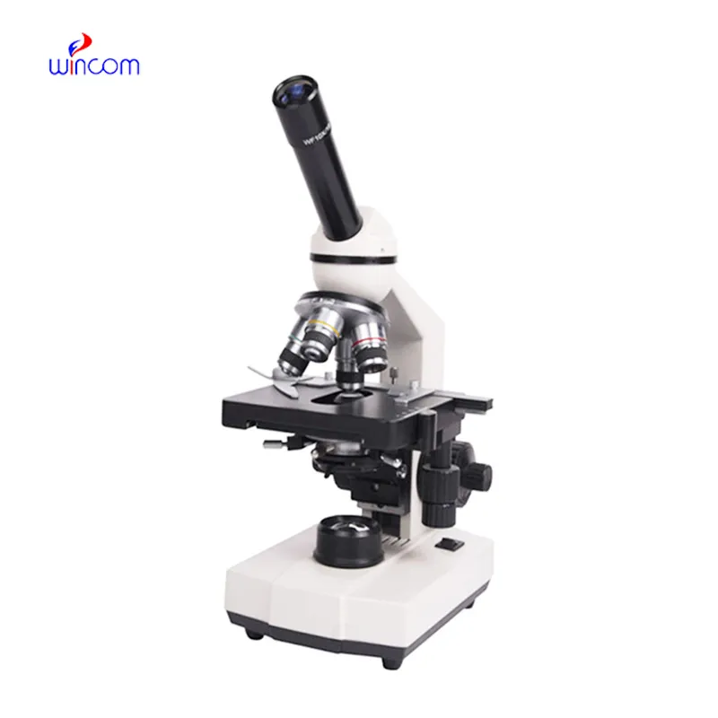
I’ve used several microscopes before, but this one stands out for its sturdy design and smooth magnification control.
To protect the privacy of our buyers, only public service email domains like Gmail, Yahoo, and MSN will be displayed. Additionally, only a limited portion of the inquiry content will be shown.
We are planning to upgrade our imaging department and would like more information on your mri machin...
I’m looking to purchase several microscopes for a research lab. Please let me know the price list ...
E-mail: [email protected]
Tel: +86-731-84176622
+86-731-84136655
Address: Rm.1507,Xinsancheng Plaza. No.58, Renmin Road(E),Changsha,Hunan,China

 af
af
 es
es
 ar
ar
 tr
tr
 sw
sw
 pt
pt
 th
th
 ur
ur
 bn
bn
 ne
ne
 vi
vi
 km
km
 lo
lo
 de
de
 ru
ru
 fi
fi
 nl
nl
 fa
fa
 fr
fr
 ko
ko