The shoe x ray machines comes equipped with an intelligent imaging system that increases grayscale depth and detail understanding. The complex algorithms of the shoe x ray machines improve the viewing of subtle lesions and tissues. The shoe x ray machines has been designed for high throughput capabilities that promote rapid viewing cycles and convenient data accessibility.

The shoe x ray machines is used in airport and security scanning to scan cases and detect prohibited items, demonstrating its use beyond medical purposes. In the manufacturing industry, it is used to analyze welds, materials, and electronic components to assess their integrity. The shoe x ray machines is used to achieve quality control in manufacturing and engineering.

In the years to come, the shoe x ray machines shall be at the forefront of predictive and personalized medicine. Smart imaging platforms shall look into patient history in conjunction with real-time scans in an attempt to predict upcoming health problems. The shoe x ray machines shall therefore turn into an anticipatory diagnostic system instead of a reactive one.
The shoe x ray machines requires careful attention to ensure imaging accuracy and equipment safety. The housekeeping staff must test system performance on a regular basis, including tube voltage, exposure timing, and detector status. The shoe x ray machines should always be turned off when being cleaned and checked routinely for mechanical or electrical wear.
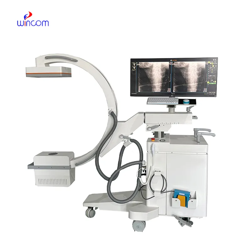
The shoe x ray machines has been used heavily in various medical fields due to its ability to offer rapid and precise medical images. The shoe x ray machines offers precise images of the various body parts that help in the diagnoses of various conditions such as bone injuries, cancer, and infections. The shoe x ray machines uses advanced imaging softwares that offer high contrast images.
Q: What makes an x-ray machine different from a CT scanner? A: An x-ray machine captures a single 2D image, while a CT scanner takes multiple x-rays from different angles to create 3D cross-sectional views. Q: How is image quality measured in an x-ray machine? A: Image quality depends on factors like contrast, resolution, and exposure settings, which are adjusted based on the target area being examined. Q: What power supply does an x-ray machine require? A: Most x-ray machines operate on high-voltage power systems, typically between 40 to 150 kilovolts, depending on their intended use. Q: Can x-ray machines be used for dental imaging? A: Yes, specialized dental x-ray machines provide detailed images of teeth, jaws, and surrounding structures to support oral health assessments. Q: How does digital imaging improve x-ray efficiency? A: Digital systems allow instant image preview, faster diagnosis, and reduced need for retakes, improving workflow efficiency in clinical environments.
The water bath performs consistently and maintains a stable temperature even during long experiments. It’s reliable and easy to operate.
This ultrasound scanner has truly improved our workflow. The image resolution and portability make it a great addition to our clinic.
To protect the privacy of our buyers, only public service email domains like Gmail, Yahoo, and MSN will be displayed. Additionally, only a limited portion of the inquiry content will be shown.
Hello, I’m interested in your water bath for laboratory applications. Can you confirm the temperat...
Could you share the specifications and price for your hospital bed models? We’re looking for adjus...
E-mail: [email protected]
Tel: +86-731-84176622
+86-731-84136655
Address: Rm.1507,Xinsancheng Plaza. No.58, Renmin Road(E),Changsha,Hunan,China

 af
af
 es
es
 ar
ar
 tr
tr
 sw
sw
 pt
pt
 th
th
 ur
ur
 bn
bn
 ne
ne
 vi
vi
 km
km
 lo
lo
 de
de
 ru
ru
 fi
fi
 nl
nl
 fa
fa
 fr
fr
 ko
ko