Using state-of-the-art real-time signal processing, the ultrasound usb scanner has the capability of producing imaging output that is invariably sharp. The system of the device is capable of dynamically tuning the frequency and gain for achieving the best image quality. The ultrasound usb scanner with its versatile probe compatibility is able to deal with different demanding clinical applications like obstetrics, cardiology, and abdominal scans.

The ultrasound usb scanner is recognized for its great contribution to the field of surgery and thus is employed frequently in operating theaters for providing intraoperative guidance and ascertaining anatomical targets. It can easily locate areas where fluid has collected, determine the condition of the tissue, and provide evidence that the procedure has been successful. The ultrasound usb scanner can also be used dynamically and thus in sports medicine for imaging of muscles and tendons during movement analysis.

The future of the ultrasound usb scanner may include enhancements based on artificial intelligence algorithms that can improve images and offer automatic measurements. Better transducer technologies may offer higher resolution and increased sensitivity. The ultrasound usb scanner may have an increasing role to play in personalized medicine because it may offer continuous monitoring capabilities.

In order to extend the service life of the ultrasound usb scanner, it is recommended that users refrain from applying much force during the process of connecting/disconnecting probes. Power cables should always remain dry. The ultrasound usb scanner needs diagnostic tests to ensure that it produces quality images.
Used in hospitals and clinics, the ultrasound usb scanner provides immediate visual feedback for a variety of medical evaluation uses. Converting sound waves into live images, the ultrasound usb scanner allows physicians to easily detect abnormalities. The ultrasound usb scanner assists with making diagnostic processes safer in addition to improving patient outcomes. It possesses an ergonomic shape alongside digital integration capabilities that support simple data sharing and medical record documentation.
Q: What imaging modes are available on the ultrasound scannert? A: It supports multiple modes such as B-mode, M-mode, and color Doppler for diverse diagnostic applications. Q: How does the ultrasound scannert improve diagnostic accuracy? A: By providing high-resolution images and real-time feedback, it enables more precise medical evaluations. Q: Can the ultrasound scannert be used in field or remote settings? A: Yes, its portable versions are designed for mobility and can be used in clinics, hospitals, or mobile healthcare units. Q: What kind of display does the ultrasound scannert use? A: It typically features a high-definition digital display that enhances image visualization and readability. Q: How is data from the ultrasound scannert managed? A: The device allows secure storage, easy access, and export of imaging data through USB or network connections.
This ultrasound scanner has truly improved our workflow. The image resolution and portability make it a great addition to our clinic.
This x-ray machine is reliable and easy to operate. Our technicians appreciate how quickly it processes scans, saving valuable time during busy patient hours.
To protect the privacy of our buyers, only public service email domains like Gmail, Yahoo, and MSN will be displayed. Additionally, only a limited portion of the inquiry content will be shown.
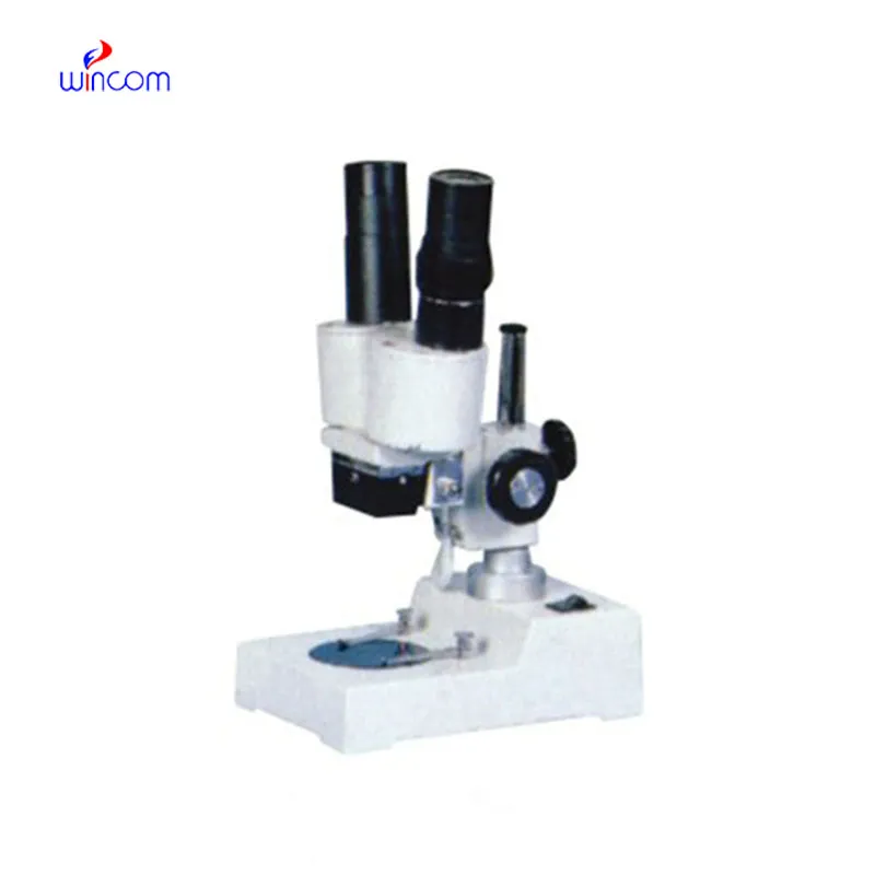
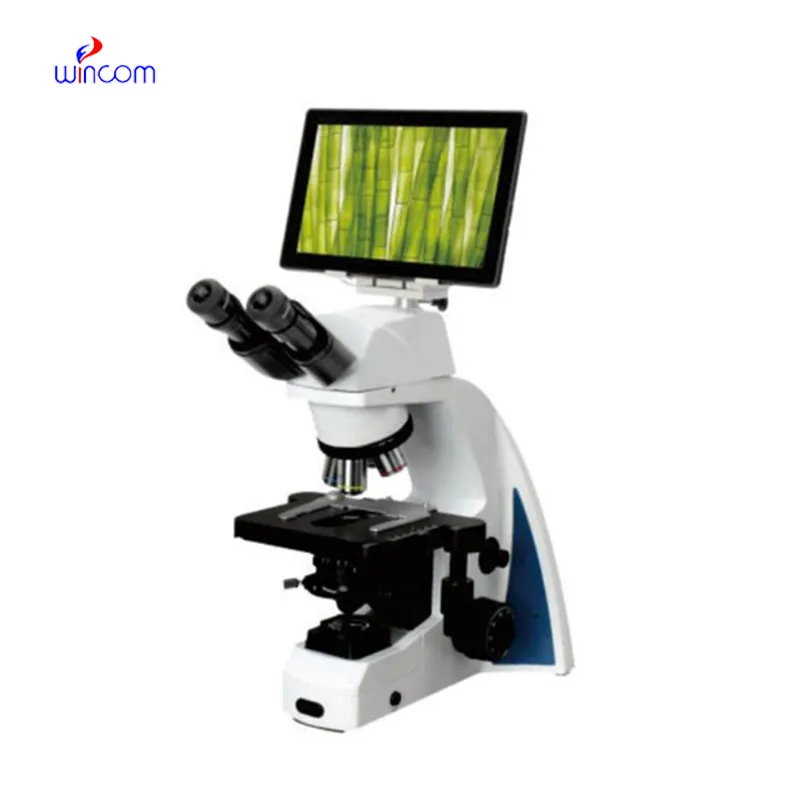
I’m looking to purchase several microscopes for a research lab. Please let me know the price list ...
Hello, I’m interested in your water bath for laboratory applications. Can you confirm the temperat...
E-mail: [email protected]
Tel: +86-731-84176622
+86-731-84136655
Address: Rm.1507,Xinsancheng Plaza. No.58, Renmin Road(E),Changsha,Hunan,China

 af
af
 es
es
 ar
ar
 tr
tr
 sw
sw
 pt
pt
 th
th
 ur
ur
 bn
bn
 ne
ne
 vi
vi
 km
km
 lo
lo
 de
de
 ru
ru
 fi
fi
 nl
nl
 fa
fa
 fr
fr
 ko
ko