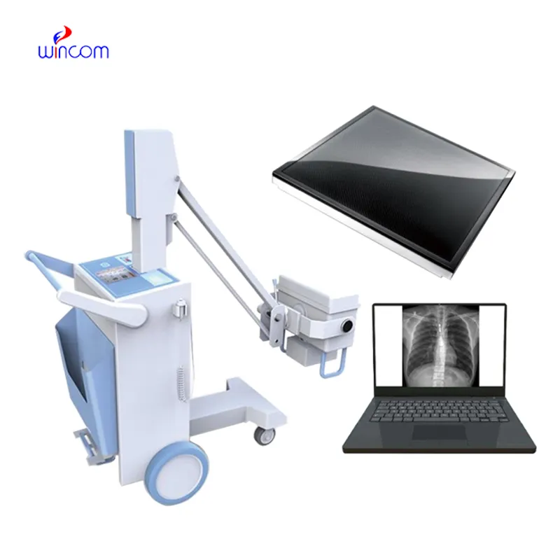
Designed with a compact and intelligent design, the x ray machine disposal offers various modes of imaging such as full body scans and localized scans. The device supports digital viewing stations where the images can be viewed remotely by radiologists. The x ray machine disposal assists in improved diagnostic operations by providing easy-to-handle mechanisms and sound imaging consistency.

The x ray machine disposal plays a central role in preventive medicine since it helps to conduct regular health screenings of the chest, spine, and abdomen. It also identifies early signs of conditions such as osteoporosis and lung disease. The x ray machine disposal allows for effective conduct of comprehensive health examinations by physicians.

With advancements in technology, the x ray machine disposal will get progressively smaller, intelligent, and networked. It will also support augmented reality for training and procedural guidance. The x ray machine disposal will have self-calibration and automated maintenance functionality, which will increase reliability and operational performance.

For the x ray machine disposal to be trustworthy, maintenance processes must include inclusive system checks, cool-down checks, and cable tests. Preventive maintenance allows potential issues to be noticed early enough before they get worse. The x ray machine disposal must be monitored for times of use and dates of inspection for traceable records of maintenance.
Through the use of high-tech detectors and digital imaging, the x ray machine disposal provides high-quality internal structural images. The device enables healthcare providers to track various conditions such as pneumonia, arthritis, and dental cavities. The x ray machine disposal offers accurate imaging and ease of handling that makes it imperative in diagnostic radiology.
Q: What is an x-ray machine used for? A: An x-ray machine is used to produce images of the internal structures of the body, helping doctors detect fractures, infections, and other medical conditions. Q: How does an x-ray machine work? A:X-ray machine emit controlled radiation that passes through the body and records varying degrees of absorption on detectors or film, creating visual images of bones and tissues. Q: Is it safe to use an x-ray machine frequently? A: Modern x-ray machines use very low doses of radiation, and protective measures such as lead aprons help minimize exposure for both patients and operators. Q: Can an x-ray machine detect soft tissue injuries? A: Although X-rays machine are primarily used to examine bones, they can reveal some soft tissue abnormalities, especially when used with contrast agents or digital image enhancement techniques. Q: Who operates an x-ray machine? A: X-ray machines are typically operated by trained radiologic technologists who ensure correct positioning, exposure settings, and safety protocols during imaging.
This ultrasound scanner has truly improved our workflow. The image resolution and portability make it a great addition to our clinic.
I’ve used several microscopes before, but this one stands out for its sturdy design and smooth magnification control.
To protect the privacy of our buyers, only public service email domains like Gmail, Yahoo, and MSN will be displayed. Additionally, only a limited portion of the inquiry content will be shown.
We’re currently sourcing an ultrasound scanner for hospital use. Please send product specification...
Hello, I’m interested in your centrifuge models for laboratory use. Could you please send me more ...
E-mail: [email protected]
Tel: +86-731-84176622
+86-731-84136655
Address: Rm.1507,Xinsancheng Plaza. No.58, Renmin Road(E),Changsha,Hunan,China

 af
af
 es
es
 ar
ar
 tr
tr
 sw
sw
 pt
pt
 th
th
 ur
ur
 bn
bn
 ne
ne
 vi
vi
 km
km
 lo
lo
 de
de
 ru
ru
 fi
fi
 nl
nl
 fa
fa
 fr
fr
 ko
ko